Professor Dr. Werner Mantele is the Professor of Biophysics at Goethe College and Frankfurt College. With over 30 years of expertise with spectroscopy, Dr. Mantele is an internationally-recognized professional within the evaluation and detection of molecules.
Inside his work, Dr. Mantele explores basic points of protein construction operate, correlation and protein evaluation by four-year remodel infrared spectroscopy. On this interview, Dr. Mantele discusses infrared spectroscopy and its makes use of in biochemistry, biology and drugs, together with its benefits, disadvantages and real-world functions.
Dr. Suja Sukumaran is a Senior Utility Scientist at Thermo Fisher Scientific. Dr. Sukumaran has over ten years of expertise in analytical instrumentation, Molecular Biology, Protein and Lipid Biochemistry, and discusses the evaluation of powdered samples utilizing an MCT detector and through multi-bounce ATR.
In ths interview they define the fundanmental points of protein structure-function coreelation and protein evaluation by Fourier Rework Infrared (FTIR) spectroscopy.
Please introduce yourselves and describe your roles at Thermo Fisher Scientific?
Dr. Werner Mantele: My identify is Professor Dr. Werner Mantele, Professor of Biophysics at Goethe College. A big portion of my work addresses the elemental points of protein construction operate, correlation and protein evaluation by four-year remodel infrared spectroscopy. I’m a Professor of Biophysics at Frankfurt College and likewise the CSO of a startup that we based six years in the past that works to get merchandise out of primary analysis on infrared spectroscopy.
Dr. Suja Sukumaran: My identify is Dr. Suja Sukumaran, the Senior Utility Scientist at Thermo Fisher Scientific. My work encompasses the above, together with using an FTIR spectrometer and microscope and presenting examples of protein evaluation.
Please define a quick historical past of Infrared Spectroscopy for us?
Dr. Werner Mantele: Infrared spectroscopy is a longtime method, which is now roughly 200 years outdated. In the event you have a look at historical past, you may see that the infrared was found round 1800: so-called ultra-red elements past the seen mild by William Herrschel, a German immigrating to London, and it step by step turned a way for molecular evaluation principally coming into chemistry.
Till the 70s, this was primarily a way utilized in chemistry labs for the identification of compounds and characterization of easy molecules. The limitation of the method has at all times been clear. However when Fourier remodeling for spectroscopy entered the sphere within the late 70s, infrared spectroscopy turned extra fascinating for biochemistry and even afterward for drugs. It developed from the primary tabletop units requiring a number of sq. meters of optical desk space to a handheld instrument that’s now in use.
A second enchancment got here across the flip of the century, specifically round 1995: the primary quantum cascade lasers as infrared sources had been developed, which was a giant step ahead as a result of it overcame some limitations of infrared spectroscopy, similar to very low energy from thermal sources. In the meantime, the quantum cascade lasers turned a dependable infrared supply.
Why did it take so lengthy earlier than infrared spectroscopy entered biochemistry, biology, and drugs?
To begin with, the spectral vary is round 2-20 micrometers. In that vary, we have now water as a powerful absorber at a number of factors of the spectrum, which makes its software in IR spectroscopy and biologically tough. You will need to remember that we have now photons within the infrared with very low vitality, nearly across the thermal vitality KT. The issue is thermal sources – heated rods, glow bars, and so forth. Thermal sources have a low brilliance, so the inherent downside of IR spectroscopy was at all times that the variety of photons was very low.
Dispersive devices require lengthy scanning occasions: the primary infrared spectra that I took as a diploma pupil within the mid-70s took hours. You can simply begin scanning a spectrum, make a espresso, have a meal, after which come again and obtain the spectrum. That turned quite a bit totally different and sooner in between.
The opposite downside was at first of IR bio spectroscopy, new sampling methods for organic macromolecules, membranes or cells had been accessible. Now, that has fully modified. To begin with, we have now compact infrared devices which work on the Fourier remodel rules, fascinating pattern interfaces, the so-called attenuated complete reflection method with simple pattern entry. These both function for very small pattern volumes, like a drop of blood, and even stream cells for bioprocess evaluation. At present, the sampling aspect can be easy and established.
Picture Credit score:Shutterstock/explode
Might you please define the second sort of infrared supply?
Dr. Werner Mantele: As I mentioned, we have now a second type of infrared supply, specifically quantum cascade lasers. These are highly effective, compact tunable mild sources with numerous photons, which get rid of one of many issues of our spectroscopy. Now we have each tunable lasers and laser arrays with numerous wavelengths, which makes spectroscopy a simple concern.
On the similar time, sampling methods have truly centered extra on organic samples. Classical sampling is a transmission pattern with easy optics and the place stream methods are attainable. Path lengths are very brief to soak up water. Sampling is a bit difficult. Cluttering can seem in case you have particles in your pattern. Definitely, this system doesn’t work in vivo. Some colleagues apply reflectance methods the place you shine infrared mild onto the pattern and gather the backscattered mild.
That is a simple method, however the penetration depth is unclear and covers only some micrometers. It’s its small pattern volumes that enable stream cells to be made. The issue is that it labored with an evanescent wave with a penetration depth usually shorter than one wavelength, so within the order of 5 micrometers, which could be limiting.
Photoacoustic methods are a lesser-known however extraordinarily handy for organic samples, particularly for working in vivo. That is the place you utilize a microphone to detect the sound wave generated by an absorption course of. That is established properly for gases and will also be used for liquids. There may be additionally a photon thermal method, which is linear and has a powerful penetration depth of roughly a tenth of a millimeter – with the chance to do depth profiling for pores and skin, for instance.
What are we going to see in infrared spectroscopy?
That is returning again to quite simple rules. Infrared spectroscopy sees adjustments of dipole moments with nuclear motions. This could reveal adjustments in bond lengths, bond angles, bond polarization of protonation reactions of salvation and the formation and breaking of hydrogen bonds. The spectral vary we have now to contemplate is from roughly two to roughly 20 microns, 5,000 to 500 wave numbers. Inside that vary, a smaller area – roughly 5 – 10 micrometers – is most related for organic samples.
What are the benefits and downsides of infrared spectroscopy?
Infrared spectroscopy makes use of small photon energies, which implies you should not have undesirable photon reactions. For instance, you aren’t prone to the injury that might occur with fluorescent spectroscopy. The photon stream is average.
In the event you design the experiment accurately, you should not have heating of the pattern. Pattern portions are additionally very small. That has been an unlimited improvement, and you don’t want excessive purity. For people who do time resolve spectroscopy, the intrinsic time decision of infrared spectroscopy is one vibration cycle, which means a number of picoseconds.
Nevertheless, there are additionally some disadvantages. As an example, infrared spectroscopy doesn’t have selectivity for particular person bonds, which means that contributions from all elements of a protein, for instance, will overlap.
As I discussed already, you’ve got a background absorption of water, which means that it’s a must to design particular pattern varieties. For instance, for proteins, the peptide bond has plenty of regular vibrations which can be properly characterised; MIA and MIB, that are the NH stretching modes. Then there’s a nomenclature which has I to VII. This numbering determines the vibration sort and amongst these, crucial one is the amide one absorbing round 16 to 700 wave numbers. That’s an almost-pure CO stretching mode.
Dr. Werner Mantele: We are going to see that that is most likely a simple option to characterize proteins. This amide one mode relies on the geometry and the secondary construction. And that data goes again to the Fifties. There’s a paper that was printed in Nature in 1950 based mostly on the construction of artificial polypeptides. This was the primary report that the NH group and the CO group of the polypeptide spine kind a construction that may be probed by infrared spectroscopy and used as a delicate probe for secondary construction.
What’s notable about secondary construction parts when examined from about 1700 to 1600 wave numbers?
Dr. Werner Mantele: In the event you have a look at a small vary in infrared spectroscopy from about 1700 to 1600 wave numbers, you’ve got secondary construction parts that take in in a different way. You discover areas the place you’ve got an unordered construction that’s on the highest. You’ve got areas the place you’ve got beta sheet absorbances, alpha-helix absorbances. And you may see that these absorbances are barely totally different in H2 and D2.
It’s clear as a result of hydrogen or deuterium is concerned there, which adjustments vibrational frequencies. In the event you analyze that Specter Vary very rigorously, you may truly get data on secondary construction.
Might you give an instance?
Dr. Werner Mantele: On a selected picture, for example, we have now a selected vary round 1610 to 1615 wave numbers, which is attribute for correct aggregated proteins or, for instance, amyloid fibrous. These are typical values; the positions of those secondary buildings differ with the size. The issue of analyzing these is that the half widths of the mode are massive, the modes are partially overlapping, and the task of transitions between secondary buildings could be tough due to that.
Nevertheless, usually talking, you’ve got an ordered construction, alpha-helix and beta sheet in several areas, and you need to use that like a fingerprint of sector construction for the characterization of the secondary construction of proteins for the affect of mutations, which can present up in that vary for protein stability and protein folding, unfolding, and misfolding.
It’s a simple, quick, and easy routine. It may be very exact if you’re searching for adjustments in secondary construction, however the issue is bends are overlapping and hardly separated.
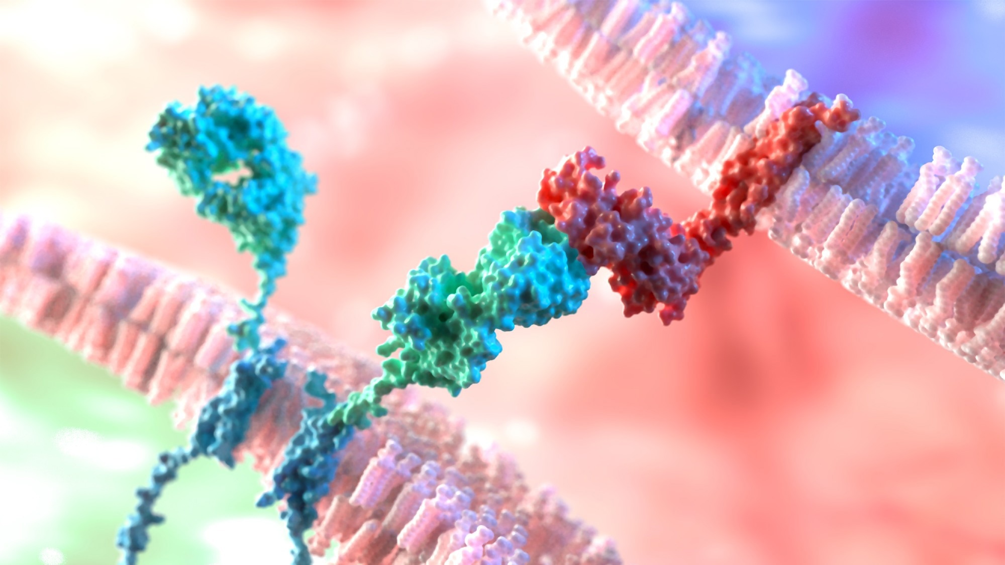
Picture Credit score:Shutterstock/AlphaTauri3DGraphics
The place does a sure secondary construction start and the place does it finish?
Dr. Werner Mantele: I cannot give an in depth evaluation of protein secretary construction, however I’ll choose up one level right here, specifically, the engaging software of direct following of protein folding. This can be a risk the place you may decide melting factors by infrared spectroscopy or see how folding and unfolding an aggregation or misfolding occur. I’ll select one small protein for this instance.
This small protein, referred to as Tendamistat, has about 74 amino acids; and eight kilos of molecular weight. This protein usually inhibits the enzyme Alpha-amylase. It has three prolines. In the event you do infrared spectroscopy in that particular vary, 1700 to 1600 wave numbers, from these proteins, and when you begin at average temperatures of roughly 20 centigrade, you see the clear sample of a beta sheet construction in keeping with the X-Ray construction of the protein. In the event you then heat up the protein to about 40, 50, 60, 70, 80 centigrade, you will note how the beta sheet sample disappears, and a transparent random coil construction seems.
The transition is about 80 centigrade. That’s totally reversible for the intact protein. In the event you settle down, the form of the curve strikes again to a transparent, beta sheet construction, in distinction to the second experiment the place we have now changed the three prolines with alanine at these positions. In the event you do the identical experiment once more, it begins with an unfolding: all of a sudden, there’s an aggregation – that is indicated by the clear sample for aggregated proteins within the infrared. This experiment is fast and provides plenty of data. In the event you do the heating up with a temperature bounce from a laser, the experiment could be accomplished in milliseconds or microseconds.
Making the swap now to biomedical functions, might you briefly define some biomedical functions of infrared spectroscopy?
Dr. Werner Mantele: The principle functions embody the evaluation of blood by infrared spectroscopy and non-invasive glucose measurement by infrared spectroscopy. That is termed biomedical spectroscopy, and there’s huge potential in that for scientific functions, moveable apps and for house use.
To begin with, the mid-infrared fingerprint of molecules in physique fluids is vital. All constituents of blood, urine, saliva, and so forth, if they’re of a molecular sort, have a transparent infrared sample within the vary of about 5 to 10 micrometers. That is the so-called fingerprint vary, and that sample can be utilized for identification and quantitative characterization – for instance, in a sensor.
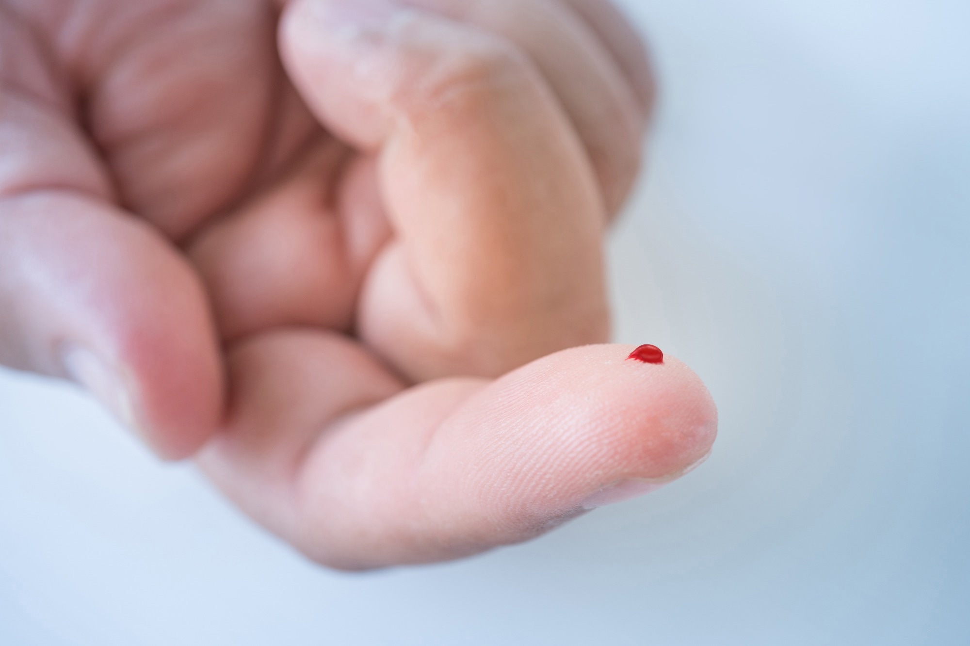
The essential precept is thus that you simply file an infrared spectrum of a blood pattern, for instance, urine samples, sweat, saliva. The evaluation takes one to 2 minutes. It’s appropriate for a degree of care and bedside functions. The pattern portions are very low, so we find yourself with a drop of blood, for instance, out of your fingertip. This technique doesn’t require any reagents; it doesn’t require a recalibration process due to reagent change. The quantity of potential infectious waste is extraordinarily low, specifically, acute tip to wipe clear the interface. It will probably use both FTIR devices or quantum cascade lasers and is developed for handheld units. The current evaluation of a double blood pattern, for instance, can yield as much as 12 medical parameters, related medical parameters.
Nevertheless, with easy modifications of the pattern interface, you too can use it for urine samples or dialysis fluid. Lastly, the prices are low: it’s simply the funding on the gadget, however there are not any consumables.
 Picture Credit score:Shutterstock/angellodeco
Might you define how the system works?
Dr. Werner Mantele: To begin with, it makes use of a compact FTIR instrument. It’s a few centimeters in measurement. Then, it wants a rigorously designed mathematical process and a rigorously chosen calibration pattern group. The consumer simply chooses the very best reference. In our case, it was the scientific lab of Frankfurt College. Within the case of full blood samples, we have now based mostly the evaluation of blood on about 2000 reference samples of full blood and plasma within the case of dialysis fluid.
The system has an computerized loading hemolysis and cleansing system. It’s tailored to scientific routines and has clinically related easy consumer software program as a result of it’s meant for medical medical doctors.
What it makes use of is mathematical procedures for calibration and validation methods. These are a number of variant evaluation, colorimetry, and even deep studying procedures. The way it works, principally, is that the consumer takes a correlation between a scientific reference worth of a sure parameter with values being within the purple vary. So, doubtlessly, the curiosity and the conventional vary, and also you plot that versus the parameter derived from infrared spectroscopy. Now, as soon as calibration has been accomplished on this manner, you’re taking an unknown pattern and also you get the clinically related worth: which, for instance, could possibly be the blood glucose worth decided noninvasively.
How can the parameters be decided?
Dr. Werner Mantele: If we stock on from the earlier query, we will state that no less than eight parameters in apply, could be decided at scientific precision. All parameters are derived inside lower than a minute for taking a spectrum and about 20 seconds’ calculation time from a single drop of blood, and they’re all at scientific precision.
These blood parameters have been investigated in a spread that covers the total scientific want, and the error is throughout the limits of what’s anticipated for laboratory chemistry, besides within the case of Immunoglobulin. This can be a platform expertise for various medical functions. Now we have examined it within the college clinics in Frankfurt for level of care evaluation in a undertaking which is named Affected person Blood Administration. Now we have a patent on urine measurements for the stream system, mainly, an ATR system with a stream cell coupled to a urinal for rapid urine evaluation. It’s used for real-time monitoring of hemodialysis for the stream cell.
Final however not least, it’s utilized for the blood evaluation of small laboratory animals as a result of the expertise requires solely a drop, whereas scientific laboratories are used for the blood evaluation of small animals. They usually want extra blood than the small animal may give with out struggling.
What are some examples of actual measurements in vivo utilizing infrared spectroscopy?
Dr. Werner Mantele: One instance which is related for many individuals on the planet at this time is its use for diabetes sufferers. In 2019, about 463 million individuals worldwide lived with diabetes, and sadly, this quantity will proceed to extend. The Worldwide Diabetes Federation estimates that by 2045, this might be near 500 million individuals. Diabetes is growing in all areas of the world: quickly in america, and quickly within the Center East. In Europe, the rise shouldn’t be as swift however is rising nonetheless and is really a world pandemic that won’t go away by itself.
What does a diabetes affected person usually do to watch or handle their illness, and the way can the portfolio at Thermo Fisher Scientific assist?
Dr. Werner Mantele: It’s well-known that diabetes can’t be cured, however the illness could be managed. Because of this a diabetes affected person measures blood glucose ranges a number of occasions a day by pricking, usually the finger, utilizing an electrochemical sensor, constructing a take a look at strip, and this sensor suits into slightly gadget. This gadget, after one or two minutes, provides the blood glucose measurement.
The issue is that these take a look at strips are expensive, particularly if you wish to do a number of measurements a day, and so they have a restricted shelf time. In the meantime, some repeatedly measuring minimally invasive sensors are tiny needles that go into your tissue and keep there for every week or two.
In fact, the problem presently is that too many individuals worldwide can not afford these steady measuring sensors, and thus, they should depend on the take a look at strips. Usually, the take a look at measurements should not frequent sufficient, and measurements happen on common about 1.5 occasions per day and no more.
Now, our idea is to make use of infrared spectroscopy. The fluid that we concentrate on shouldn’t be blood, however it’s interstitial fluid or pores and skin fluid. It’s a liquid that surrounds cells, pores and skin and muscle, and it’s greater than a minimal quantity. It’s twice the amount of blood. That liquid usually seems on the pores and skin after shallow scratches or seems in a modified blister. It’s a matrix of water ions, albumin, glucose, and phosphate.
Glucose in interstitial fluid represents about 90% of blood glucose, and if blood glucose adjustments, the ups and downs of blood glucose present up with little or no delay in interstitial fluid. Our portfolio permits for the focusing on of interstitial fluid within the pores and skin with spectroscopy.
 Picture Credit score:Shutterstock/NatachaS
How does focusing on of interstitial fluid within the pores and skin with spectroscopy enable for a non-invasive glucose measurement?
Dr. Werner Mantele: To begin with, we use the infrared fingerprint of glucose. These are glucose spectra in water at concentrations related for diabetes sufferers. The decrease restrict corresponds to 50 milligrams per deciliter, which is hypoglycemic for diabetes sufferers. The subsequent restrict is 100 milligrams per deciliter, which is regular glucose. And the highest restrict is 500 milligrams per deciliter for hyperglycemic conditions, which can be of curiosity for diabetes sufferers. These peaks come up from CO and OH bonds of the glucose molecule. This sample is so attribute that it may be termed a glucose fingerprint within the mid-infrared.
We make use of that fingerprint within the infrared utilizing a laser system. You must ship infrared mild into the tissue. Infrared radiation at 10 micrometers penetrates about 60 to 100 micrometers. We should not have the chance to make use of transmission measurements, however we will use photothermal or photoacoustic applied sciences to detect the absorbance course of right here.
How are photothermal or photoacoustic applied sciences used to detect the absorbance course of?
Dr. Werner Mantele: In our setup, we use an inner reflection component. We use an infrared beam – referred to as a pump beam – from a quantum cascade laser that shines lights by way of this crystal into pores and skin. The beam penetrates about 100 micrometers of pores and skin. If you choose the glucose-specific wavelengths for this pump beam, glucose molecules within the pores and skin’s interstitial fluid will take in the infrared beam. As a consequence of absorption, they deposit a tiny quantity of warmth in these layers, that are warmed up by absorption simply by a number of millikelvin. The consumer doesn’t really feel it.
Now, this warming up of the pores and skin layers at that depth reveals up on the floor and is transferred into that inner reflection component.
What occurs within the inner reflection component?
Dr. Werner Mantele: This tiny warming up generates a so-called short-term thermal lens. A brief thermal lens is one thing that you realize from on a regular basis expertise. In the event you drive within the summertime on an asphalt highway, and the solar burns on the asphalt, you see that the air instantly in touch with the asphalt has totally different optical properties. It adjustments the optics, and you may even see a mirage impact due to that. And that’s precisely a thermal lens.
We use a second laser beam, which is named ProBeam, which is an easy purple laser diode that we ship by way of the thermal lens. It’s deflected on the thermal lens, and the deflection is detected by a place that’s delicate to the diode.
This deflection is proportional to the absorbed radiation to the pump laser energy, which we will management, and it’s depending on the change of the refractive index with the temperature of the inner reflection component. It’s IR detection by seen mild, which permits for the non-invasive measurement of glucose.
It’s attainable to make use of infrared spectroscopy for blood glucose measurement with none invasive process. It’s exact sufficient for use by diabetes sufferers. Its accuracy corresponds to commercially accessible glucometers. That was printed each final 12 months and this 12 months within the Journal of Diabetes Science and Expertise.
Suja, might you please present an overview of your work concerning experimental points of protein evaluation?
Dr. Suja Sukumaran: My work focuses on the evaluation of proteins in FTIR, in addition to establishing how protein evaluation can be utilized for functions in meals, expertise, and analysis. We’d like to consider protein construction, stability, and aggregation. In analyzing samples in FTIR, there are two key sampling methods that we have now to recollect. One is transmission, for which we will use transmission cells that are very small path hyperlinks. We will additionally use protein options which can be in H2O or D2O-based buffers. What we are going to do at this time is look ATR, and we are going to use each multi-bounce ATR in addition to single-bounce ATR.
Multi-bounce ATR – in addition to the single-bounce ATRs – is likely one of the extra in style strategies for evaluation these days due to its ease of use, ease of cleansing, and the chance to regain the pattern after the experiment is accomplished.
 Picture Credit score:Shutterstock/TatjanaBaibakova
Which detector ought to be utilized in this sort of experiment?
Within the scenario outlined under, an MCT detector ought to be used, particularly if you’re working with proteins at very low concentrations.
An MCT detector, which is a liquid nitrogen cooled detector, is far more delicate than the DTGS detector and far sooner as properly. There are, subsequently, choices for the consumer to make use of the MCT and DTGS. The gather tab will inform the place we will set the parameters for the variety of scans and the decision to different key parameters, which is predicated on what samples are being measured and what finish aim you’ve got. In case you are contemplating samples which can be very dilute, you’ll want to go a lot greater in scan numbers.
Why is the decision parameter necessary?
Dr. Suja Sukumaran: The decision parameter is necessary as a result of if you’re planning on doing a secondary construction analysis or have a look at peak shifts and peaks which can be very shut to one another, and also you need to resolve them correctly, you might need to use a decision of two. For common use, you too can use a decision of 4. For greater resolutions, the time required to make a set can even be greater. The variety of scans can be vital right here as a result of there might be experiments the place you’re searching for very small variations between the identical protein pattern. You undoubtedly need to go greater within the variety of scans in order that your sign to noise is actually good.
How is the background measured?
Dr. Suja Sukumaran: Inside the OMNIC software program, the consumer can simply click on on this button that claims “Click on Background,” and it’ll begin the background measurement. As soon as the background measurement is accomplished, the consumer is prepared for both their pattern or the buffer measurements simply by themselves. All of the consumer wants is ten microliters of my buffer or the pattern. To start out the measurement, merely click on “Acquire Pattern.”
Relating to the measurement of buffer and protein, the subsequent step can be to subtract the buffer. To subtract the buffer, all we have now to do is click on on the “Choose all” button after which, below “Course of”, click on on “Subtract”.
Now, we have now to make use of the amide I area for our peak decision or for peak becoming to get the second to restructure.
For that, we simply choose the amide I area. And all we have now to do is go to the analyze button and click on on peak resolve. In peak resolve, as soon as we have now chosen the peaks and we have now described the noise ranges, all we have to do is click on on the thick peaks button. This may run fairly a number of iterations to suit the peaks into the spectrum by itself. As soon as the height becoming is completed, all we have to do is click on on the button in order that it simply provides it into a brand new window.
We will additionally have a look at the peaks and the height areas as properly. By simply trying on the peaks, you can begin to see the varied peak areas that the software program has give you. You’ll be able to add and delete sure peaks as and when required.
How can the proteins be analyzed utilizing a multi bounce ATR?
Once we need to analyze proteins which can be in water-based buffers, we have to gather a buffer spectrum. We have to gather the protein spectrum, particularly if we’re going to do that at totally different temperatures additionally; we undoubtedly should gather the buffers at totally different temperatures, in addition to the protein at totally different temperatures.
Then, we subtract the buffer from the protein spectrum, after which the subtracted protein spectra with the amide I area between 1,700 to 1,600 is used for the height consequence or the height becoming operate.
Extra data could be obtained below the Omnic coaching movies on our web site.
There’s a second option to analyze this and that’s utilizing the BioTools PROTA-3S software program. Utilizing the software program, you are able to do a number of spectra on the similar time, however this software program can be extra geared in the direction of transmission evaluation. If in case you have samples the place you’re doing a number of samples in transmission, you need to use the PROTA-3S software program for evaluation of secondary construction as properly.
Are you able to speak us by way of the applicability of those options?
One of the crucial commonplace examples of this type of protein secondary construction dedication is once we take into consideration powdered meals. Whey, rice, milk protein and pea protein are all supplies which can be made up of complicated mixtures of carbohydrates, proteins, and lipids. Nevertheless, it’s easy and simple to research them: this may be accomplished in a single bounce ATR.
It is vitally highly effective to make use of a PCA-based evaluation of those powders for classification and discrimination evaluation.
When I’ve the pattern, all I must do is take a small scoop of it and put it on the ATR crystal (assuming the background has been accomplished). After getting a powdered pattern or so, you will need to use the strain tower that’s proper in there in order that the pattern is in correct contact with the ATR crystal by itself. Then merely click on “Acquire Pattern,” and the assortment begins.
What could be noticed within the evaluation of the outcomes from related powdered samples?
Now we have analyzed milk protein, pea protein, rice protein, and whey protein from totally different distributors, in addition to from totally different tons that had been analyzed, and so they had been analyzed repeatedly.
Upon inspection, even the identical sort of protein from totally different distributors confirmed delicate variations. The spectrum was then additional processed utilizing PCA evaluation. What we noticed was that every of the proteins would separate out into its personal little precept part areas.
The benefit of that is that the subsequent time a sure sort of protein pattern from a selected vendor arrives, they will examine it to this explicit mannequin and say, “Oh, so this one is way nearer to the G1 group” or “That is similar to the milk protein group that we noticed within the A2 sort”, or so on.
What’s the Rheonaut, and the way is rheology associated to meals and drinks?
A Rheonaut is an instrument the place rheology is coupled with FTIR spectroscopy. We need to find out about viscosity and in regards to the elasticity of the fabric. We need to know in regards to the viscosity of issues like chocolate syrup, in addition to the elasticity of supplies like marshmallows.
We knew we wanted to couple two methods: the primary learning the bodily properties of supplies, like meals powders, particularly proteins, after which the second is the FTIR spectroscopy. That resulted within the instrument referred to as the Rheonaut.
If we have a look at the Rheonaut, it’s made up of three primary elements. One is the FTIR, the opposite one is the HAAKE MARS rheometer by itself, after which there’s the interface between the FITR and the rheometer. Inside this interface, the important thing part is the diamond ATR, which is constructed below the metal plate of the rheometer by itself.
How does the consumer load the pattern into the metal plate?
The whey protein, for example, is loaded proper into the metal pedestal. Beneath, there’s additionally the diamond ATR crystal. The RheoWin software program controls the temperature ramps, nevertheless it additionally communicates with the Omnic software program. On this maneuver, we’re gathering rheological property and the adjustments within the FTIR peaks utilizing the FTIR. The temperatures are managed proper on the pedestal when the plates make contact.
When the temperature ramps up and cools down is over. On the finish of the experiment, the liquid materials we loaded in has turn out to be this good gel-like materials that can be versatile and might fold. On the similar time, there are important adjustments within the amide I and II peaks, which signifies that there’s some aggregation of protein that has occurred as there’s a downshift within the peak place, in addition to a change within the depth of the fabric.
While you truly start within the resolution state, the whey protein is in a pleasant folded state. It unfolds because the temperature ramps up, leading to aggregation and the event of the entire gelation course of.
What had been the necessary findings from this research?
We will use protein secondary construction, folding and aggregation research for a lot of totally different software varieties, and we will carry out these utilizing transmission.
Lastly, we noticed that we might do that with ATR: each single bounce and multi bounce ATR. We additionally discovered that we might use the ability of FTIR spectroscopy and mix it with PCA evaluation to create some actually highly effective discriminate fashions to do some good QC methods. We will additionally use hyphenated methods, just like the Rheo-IR, and different issues just like the TGA-IR or GC-IR to know numerous bodily and chemical properties of on a regular basis supplies.
About Werner Mäntele, PhD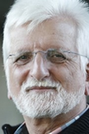
Prof. Dr. Werner Mäntele is a professor of biophysics on the Goethe College in Frankfurt am Predominant. He has over 30 years of expertise with spectroscopy and is an acknowledged professional in analyzing and detecting molecules like glucose and protein. At the moment, Prof. Dr. Mäntele is the chief scientific officer in DiaMonTech, a medical gadget firm based mostly on the idea of “photothermal detection”, which Prof. Dr. Mäntele and his staff developed.
About Suja Sukumaran, PhD
Dr. Suja Sukumaran earned her PhD in Biophysics from Johann Wolfgang Goethe College, Germany, as a part of the worldwide Max Planck analysis Faculty. She is the co-inventor of US PATENTS on ‘MspA Nanopores and associated strategies’ licensed to Illumina Inc. Her expertise and experience is in Molecular Spectroscopy, Seen and fluorescence Imaging, protein and lipid biochemistry. Her present analysis pursuits are AI for protein folding , microplastics and recycling.
About Thermo Fisher Scientific – Supplies & Structural Evaluation
 Thermo Fisher Supplies and Structural Evaluation merchandise offer you excellent capabilities in supplies science analysis and improvement. Driving innovation and productiveness, their portfolio of scientific devices allow the design, characterization and lab-to-production scale of supplies used all through business.

